Other special stains
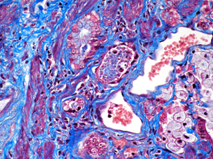
Masson staining
Two or three anionic dyes are mixed together or act successively to complete the dyeing. According to the different permeability of tissues, anionic dyes with different molecular sizes are selected for dyeing, and different tissue components can be displayed. It is mainly used for the differential dyeing of collagen fibers and muscle fibers. Masson staining is the most commonly used connective tissue staining method. This method is mostly used to observe the proliferation and distribution of fibrous connective tissue in diseased tissues and to distinguish fibrous tumors from myogenic tumors. It has important value for the difference of fibrous tissue proliferation in lobular portal area caused by alcoholic cirrhosis / post necrotic cirrhosis and various types of hepatitis.
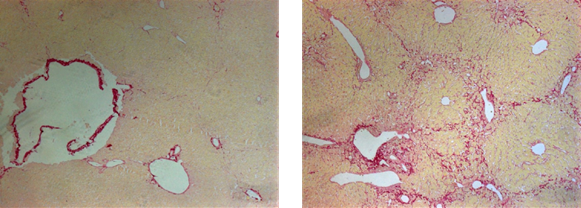
Sirius red staining
Sirius red and its lining dye solution are strong acid dyes, which are easy to combine with the basic groups in collagen molecules and adsorb firmly. Polariscope showed that the collagen fibers had the property of positive uniaxial birefringence. After combined with Sirius red staining solution, it could enhance the birefringence and improve the resolution, so as to distinguish the two types of collagen fibers. Collagen fiber is the most widely distributed and abundant fiber in connective tissue. It is widely distributed in various organs, including skin, sclera and tendon. The solvent of Sirius red staining solution is PA saturated solution. It is mainly used in the study of collagen fiber abnormalities or fiber proliferation in various tissue lesions. The collagen fibers of tissues such as tubes are dyed red to help.
Oil red O staining
Oil red O is an azo dye. It is a strong lipid solvent and dye. It combines with triglycerides in the form of small lipid droplets. Fat soluble dyes can be dissolved in lipids in tissues and cells, and its solubility in lipids is greater than that in solvents. When the tissue section is filled with dye, the dye leaves the dye and dissolves in the lipids (such as lipid droplets) in the tissue, making the lipid droplets in the tissue orange.
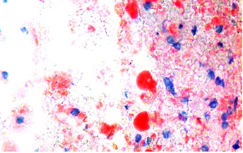
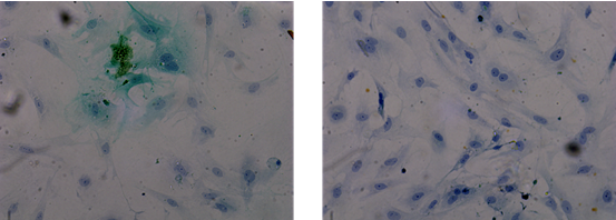
Toluidine blue staining
Toluidine blue is a kind of commonly used synthetic dyes, belonging to quinone imine dyes. This kind of dyes generally contain two chromophores, one is amino group, the other is quinone benzene ring; Toluidine blue contains not only two chromophores, but also two auxochromes. It is an alkaline dye. The cations in toluidine blue have dyeing effect, and the acidic substances of tissues and cells are dyed by combining with the cations, staining the nucleus to make it blue; The cytoplasm of mast cells contains heterochromatic substances such as heparin and histamine, which can be metachromatic purplish red when encountering toluidine blue. It can be used for the preliminary screening of condyloma acuminatum and the detection of mast cells.
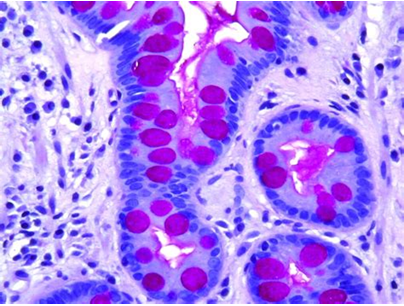
PAS staining
PAS staining is also known as periodic acid-Schiff staining and glycogen staining. It is generally used to detect sugars in tissues. Periodate can oxidize the polysaccharide glycol group in the cell to dialdehyde, and then combine with the colorless magenta of Schiff's solution, which is red and located in the cytoplasm.
Giemsa staining
Giemsa dye is composed of azurol and eosin. The dyeing principle and results are basically the same as those of wright staining. Eosinophilic particles are basic proteins, which are combined with eosin and dyed pink, which are called eosinophilic substances; Nuclear protein and lymphocyte cytoplasm are acidic, combined with alkaline dye methylene blue or azure, dyed purple blue, which is called basophilic substance; Neutral particles are in an isoelectric state and can be combined with eosin and methylene blue. They are dyed lavender and are called neutral substances.
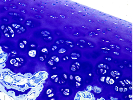
Nissl staining
Nissl staining is a method of dyeing nerve tissue with alkaline dye. Nissl body is a kind of basophilic substance in cytoplasm, which is widely seen in various neurons. The shape, size and quantity of Nissl body in different neurons are different. The basic dyes used in Nissl dyeing mainly include tar violet, methylene blue, toluidine blue and thionine. Our room is mainly stained with tar violet and thionine. Nissl staining can stain Nissl bodies to observe the cell structure in neurons; The damage of neurons can also be understood through the observation of Nissl body after Nissl staining.
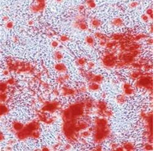
Alizarin red staining
Alizarin red staining forms a complex with calcium ions to identify the calcium salt components in tissues and cells. The process of osteogenic induction is to enable calcium ions to precipitate in the form of calcium salt, namely "calcium nodules". Alizarin red staining is commonly used to identify calcium nodules. Osteogenic differentiation is an important characteristic of stem cells. After a specific induction medium for a period of time, stem cells can differentiate into osteoblasts, deposit calcium salts on the cell surface and form calcium nodules. The complex dye of alizarin red can be used to identify whether stem cells have successfully transformed into osteoblasts.
TTC staining
2,3,5-triphenyltetrazolium chloride (TTC) is a fat soluble light sensitive complex, which is often used to detect the viability of seeds and the ischemic infarction of mammalian tissues. It is a proton receptor of pyridine nucleoside structural enzyme system in respiratory chain. It can react with dehydrogenase in normal tissue and turn red. The dehydrogenase in the living cells of normal brain tissue and viable seed embryo tissue can reduce TTC to insoluble red stable triphenylmethyl (TTF). If the brain tissue cells or embryo cells die or decline in viability, they cannot be stained or the staining is shallow. Therefore, the activity of brain tissue or seed can be identified according to the staining site and the depth of staining.
Results: the normal tissue was stained red, and the cerebral infarction area was not stained.
|