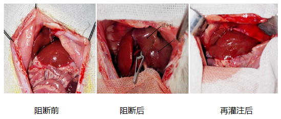
Liver ischemia-reperfusion model
Ischemia reperfusion injury is a common tissue and organ injury in clinical surgery, playing an important role in the pathological evolution of severe infections, trauma, shock, cardiopulmonary dysfunction, and other diseases. The pathogenesis of ischemia-reperfusion injury in the body is currently mainly believed to be related to the generation of a large number of free radicals in the body during ischemia, which leads to lipid peroxidation of tissues and organs associated with ischemia. Blocking and reperfusion of the portal vein and hepatic artery in the middle and left lobes of the liver in anesthetized animals can cause significant reperfusion injury due to the blockage and reperfusion of blood flow in the middle and left lobes of the liver.
Liver ischemia-reperfusion rat model
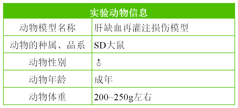
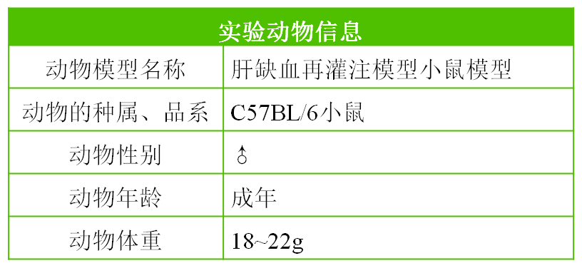
Liver ischemia-reperfusion mouse model
Observation indicators
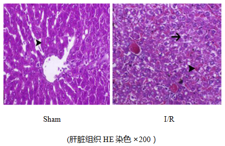
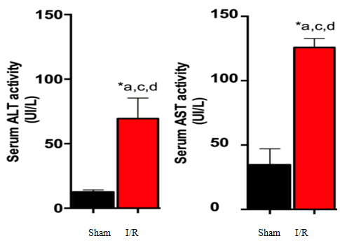
Pathological examination of liver tissue shows that the structure of liver lobules can still be seen in the liver tissue, but liver cells show watery degeneration, and the cytoplasm is loose and vacuolar; Some liver cell nuclei are heavily stained and have undergone pyknosis and reduced volume; Small focal necrosis can be seen in the liver tissue, and the liver cell structure within the necrotic lesion has disappeared into an empty network. Only residual cell fragments and inflammatory cell infiltration dominated by neutrophils can be seen; Upregulation of serum AST and GST levels, increased MDA activity and inflammatory factors, and decreased SDA activity.
Partial experimental results display
